Table of Contents
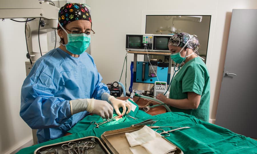
Whether you use single-function or multi-parameter devices, accurately interpreting the data provided plays a critical role in monitoring the anesthetized patient.
Hands-on monitoring
Despite the availability of more technologically-advanced options, hands-on monitoring remains a method of choice for many animal health professionals due to its simplicity and effectiveness.
Anesthetic depth is typically measured using reflexes. When a patient is under a deep plane of anesthesia, the palpebral reflex should be absent, but the corneal reflex should remain present. Loss of the corneal reflex indicates that the patient is too deep and in danger. Jaw tone can also be used throughout anesthesia to assess anesthetic depth, as can the presence or absence of a withdrawal reflex (present only under a light plane of anesthesia).
Cardiovascular status is typically assessed using mucous membrane color and capillary refill time. These observations can also be affected by drugs, but they are still a useful means of assessing the patient’s cardiovascular status.
Warning signs
Increases in capillary refill time may indicate hypovolemia, hypotension, or poor tissue perfusion. These observations should be confirmed using other objective monitors, but can often be the first indicator to the anesthetist that there is a problem warranting investigation.
Changes in mucous membrane color may have a variety of causes. Mucous membrane color may become pale in cases of vasoconstriction, hypotension, hypovolemia (hemorrhage), or hypoxia. Mucous membrane color may become dark pink in the presence of vasodilation, hypercarbia, or toxic changes. Hypoxemia may lead to blue or purple mucous membranes.
Peripheral pulses should also be regularly palpated in anesthetized patients. Weak or bounding pulses may provide an early indication of a problem that requires investigation. These observations should always be confirmed using blood pressure measurements, but can provide a quick and easy means for assessing perfusion.
The heart should also be auscultated regularly in anesthetized patients, if possible. Although surgical drapes may not always make this practical, auscultation allows for the detection of heart murmurs, abnormal lung sounds, and other abnormalities that cannot be detected via other monitors.
Pulse oximetry
Pulse oximetry is used to assess the patient’s oxygenation status. The pulse oximeter measures differences in light absorption between oxygenated and unoxygenated blood. The measurement provided by a pulse oximeter is the SpO2, which estimates the percent oxygenation of the arterial blood.
A pulse oximeter can be placed on any part of the body with adequate blood flow; common sites include the tongue, ear, vulva, prepuce, or toes. In some cases, flat probes may be used that can be placed in the rectum or esophagus, or against a bony structure such as the base of the tail.
Pulse oximetry is common in most practice settings, because it is inexpensive and easy to perform. However, the accuracy of pulse oximetry readings decreases in the presence of patient movement, poor circulation, anemia, low body temperature, and can be affected by ambient light, drugs used, skin pigmentation, and probe placement.
Capnography
Capnography measures end-tidal CO2 (ETCO2), which is the amount of carbon dioxide present in air exhaled by the patient. Exhaled carbon dioxide levels reflect blood levels of carbon dioxide, so this measurement allows the patient’s ventilatory status to be monitored.
Abnormally low carbon dioxide levels indicate hyperventilation, while abnormally high carbon dioxide levels indicate hypoventilation (inadequate breathing). A capnograph is especially useful in determining the patient’s respiratory rate when chest movement or movement of the anesthetic bag is difficult to see.
Capnography readings can also provide the first signs of problems in the anesthetic breathing circuit. Abnormal capnograph waveforms may indicate a closed valve in the anesthetic circuit, while abnormally high inspired CO2 levels may indicate exhausted CO2 absorbent.
Blood pressure
Blood pressure monitoring is used to assess pressure within the circulatory system and, therefore, organ and tissue perfusion. Low blood pressure indicates inadequate blood flow to the organs, which can lead to organ failure.
Blood pressure can be measured by one of three methods:
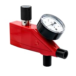
1. Doppler flow detector monitoring (Indirect)
A Doppler monitor makes sound as arterial blood flow passes by the Doppler probe. A blood pressure cuff is used to occlude the artery by inflating the cuff, and the sounds produced by the Doppler are used to determine whether blood is flowing past the cuff at a given pressure.
Doppler monitors are very accurate on small patients and can provide a continuous assessment of heart rate. Disadvantages of using a Doppler monitor include the need to clip hair from the measurement area and the fact that placing a Doppler correctly requires training.
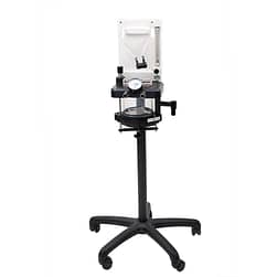
2. Oscillometric blood pressure monitor (Indirect)
This is another non-invasive technique for measuring blood pressure. A monitor is used to electronically detect arterial oscillation within an inflated cuff placed around the patient’s limb or tail. This allows the patient’s systolic, diastolic, and mean arterial pressures to be calculated and displayed on the monitor.
3. Invasive blood pressure monitoring (Direct)
This technique is typically reserved for critically-ill patients. A catheter must be placed directly into an artery and attached to a transducer via saline-filled tubing. Although it is considered to be the most accurate, this technique is invasive and requires specialized skills and veterinary anesthesia equipment.


Electrocardiogram
ECG monitoring provides valuable information about electrical impulse conduction within the heart. In anesthetized patients, ECG monitoring can allow the detection of drug-induced arrhythmias, as well as arrhythmias that may be caused by electrolyte abnormalities.
It is important to note that an ECG only confirms electrical activity. A patient can be dead and still have electrical impulses on ECG; an ECG cannot confirm that the heart is actually beating and moving blood.
Three ECG leads are typically applied to anesthetized patients: one on each of the front legs, and one on the left rear leg. Alligator clips are typically applied to the skin and then wet with isopropyl alcohol.
Body temperature
Patients generally lose body heat while under anesthesia. This can be counteracted using a variety of warning devices, such as warm water circulating blankets or warm air convection blankets.
While less common, hyperthermia may also be observed. This may develop as a reaction to anesthetic drugs, or in the case of patients who are febrile before surgery.
Temperature should be monitored at least every 15 minutes, if not more frequently.
Anesthesia monitoring cheat sheet
| Observable Parameter | Normal Values |
|---|---|
| Palpebral reflex | Present under a light plane of anesthesia; absent under a deep plane of anesthesia |
| Corneal reflex | Should be present; absence indicates excessively deep anesthetic plane with high risk to patient |
| Jaw tone | Varies with anesthetic depth |
| Capillary refill time | ≤ 2 seconds |
| Mucous membrane color | Pink |
| Peripheral pulses | Strong, synchronous |
| Pulse oximetry | SpO2 > 95% |
| Capnography | ETCO2 35–45 mm Hg |
| Blood pressure | Mean 70–120 mm Hg (most important measurement, because it is most closely correlated to organ perfusion) Systolic 100–140 mm Hg; Diastolic 50–100 mm Hg |
| ECG | Normal sinus rhythm or respiratory sinus arrhythmia |
| Body temperature | 97-101° F (36.1 – 38.3° C) |
FAQs in anesthetic monitoring
Does wetting the pulse oximeter help it work more effectively?
No. Many veterinary team members wet the tongue in order to help improve pulse oximetry readings, but there is no evidence that this is effective and no reason that it should improve values.
More likely, removing and replacing the probe allows for improved blood flow into that area of the tongue and this is responsible for the improved readings.
How do I choose the correct oscillometric blood pressure cuff for my patient?
When sized correctly, the width of the blood pressure cuff should be 40% of the limb circumference in dogs and 30% of the limb circumference in cats. Using an appropriately-sized cuff is essential in order to obtain accurate measurements.
Where should a blood pressure cuff be placed on a veterinary patient?
When recording blood pressure, the measurement must be taken at heart level. Therefore, the patient should be in sternal or lateral recumbency. If you are using oscillometric blood pressure monitoring, place the cuff distal to the elbow on the forelimb or around the mid-metatarsus on the hindlimb.
If you are using Doppler monitoring, you should place the cuff mid-radius on the forelimb and proximal to the hock on the hindlimb. (The transducer is placed over the peripheral artery on the palmar or plantar surface).
Sources and additional reading
- Baetge, Courtney. 2007. Back to Basics: “Monitoring the Anesthetized Patient.” Veterinary Technician. Vol 28, No 1. Accessed at: http://www.vetfolio.com/monitoring-and-nursing/back-to-basics-monitoring-the-anesthetized-patient
- Bryant, Susan. 2011. Facts and fiction about pulse oximetry. Presented at Central Veterinary Conference, San Diego. Accessed at: http://veterinarycalendar.dvm360.com/facts-and-fiction-about-pulse-oximetry-proceedings
- Durham, Edward. 2005. Arterial Blood Pressure Measurement. Veterinary Technician. Vol 26, No 5. Accessed at: http://www.vetfolio.com/monitoring-and-nursing/arterial-blood-pressure-measurement
- Lafferty, Katrina. 2016. Bells and Whistles: Ins and Outs of Anesthesia Monitors. Presented at Wild West Veterinary Conference.
About the author


