Canine castration, or dog neutering, is a simple surgery that small animal veterinarians perform on a daily basis. Although complications are relatively uncommon, a solid understanding of surgical technique and predisposing factors is required to ensure the best possible outcome.
Table of Contents
Surgical technique
A routine dog neuter (in a dog with both testicles descended) is generally performed through a prescrotal incision. This incision is made just cranial to the scrotum and extended to a length that is sufficient to allow exteriorization of the testicles.
Another less common castration technique is performed via a single scrotal incision, near the median raphe. This typically decreases surgical time. Although scrotal incision is a less common surgical approach among general practitioners, it has been described as a safe and efficient approach in both pediatric and adult dogs. The complication rates between the two techniques are also relatively similar.
Once the skin incision has been created, regardless of whether it is in a scrotal or prescrotal location, the surgeon must select between an open and closed castration technique.
In a closed castration, the fascia is stripped from the spermatic cord to allow the testicle to be exteriorized. The parietal tunic is left intact. Two hemostats are used to crush the testicular cord proximal to the testicle. Sutures are then placed and the testicular cord is transected distal to the hemostats. The hemostats are removed, the testicular cord is checked for bleeding, and the testicular cord is returned to the abdomen. The same procedure is then repeated for the other testicle.
In an open castration, the parietal tunic is incised to allow visualization of the vas deferens and spermatic vessels. The vas deferens and spermatic vessels may be ligated together or separately, depending upon the size of the dog. Some veterinarians feel that this technique is safer than a closed castration because there is less risk of ligature slippage, but there is little controlled research to support this view.
Intraoperative risks
Unlike ovariohysterectomy, the risk of significant intraoperative surgical complications during a routine (non-cryptorchid) dog neuter is relatively low. Slipped pedicles, nicked spleens, and other intraoperative risks associated with ovariohysterectomy are uncommon during a routine castration and also relatively uncommon during cryptorchid castration.
The greatest intraoperative risk during a routine dog neuter lies in the level of risk that is inherent in anesthesia.
A number of studies have examined the risk of significant complications or mortality in pets undergoing anesthesia. A 1998 study found that 2.1% of anesthetized dogs will experience some sort of anesthetic complication, with an anesthetic mortality rate of 0.11%.
Although the risks associated with anesthesia are low, steps should be taken to minimize these risks to the extent that is possible. Elective procedures such as neutering should be reserved for healthy pets, with steps taken to correct illness prior to anesthesia. Proper equipment and safe anesthetic protocols should be utilized, with customized protocols designed for each patient using a balanced anesthetic approach.
Anesthetic monitoring, such pulse oximetry, blood pressure measurement, capnography, and electocardiography should be utilized for all anesthetized patients. This monitoring can allow for the early detection and correction of anesthesia-associated problems.
Post-surgical complications
Estimates of complication rates associated with routine canine neutering range from 0 to 32%, with younger patients often associated with higher complication rates. Many complications likely go undetected, as owners probably monitor mild complications at home without seeking veterinary care.
Commonly-reported complications of dog neutering include the following:
- Dehiscence of the surgical incision
- Scrotal hematoma
- Bruising
- Hemorrhage
Many complications are also associated with self-trauma to the surgical site; this may be either a cause or an effect of the complications listed previously.
Surgical site dehiscence
Most cases of surgical site dehiscence are caused by self-trauma. The risk of self-trauma, and therefore dehiscence, can be minimized with appropriate surgical technique.
Avoid suturing too tightly when closing the incision, as tight sutures may lead to pain, discomfort, and an increased risk of self-trauma.
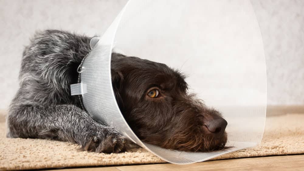
Additionally, an Elizabethan collar (e-collar) may be used during the patient’s recovery to prevent self-trauma to the incision.
Scrotal hematoma
In many cases, scrotal hematomas are a relatively mild complication that will resolve with time. In some patients, however, scrotal hematomas may become large enough to compromise blood flow within the scrotum. These patients may develop necrosis of the scrotal skin, ultimately requiring scrotal ablation for treatment.
Scrotal hematoma may be caused by bleeding from the testicular artery, or bleeding within the scrotum. At the first sign of a hematoma forming, ice packs and non-steroidal anti-inflammatory medications can be used to slow the progression of this condition.
Bruising
Capillary bleeding may be noted in the initial hours post-surgery. If this occurs, the use of a light scrotal wrap to apply pressure to the incision can help control this bleeding. This wrap can be removed after a couple of hours. Doing this can help minimize post-surgical bruising at the incision.
Hemorrhage
Hemorrhage following a dog neuter is typically caused by ligatures that have slipped due to insecure placement. There are a number of techniques that may be used to decrease the likelihood of this complication, including:
- Using a Miller’s knot for ligation of the testicular cord
- Double-ligation of the testicular cord, using a transfixation suture as the distal suture
If significant bleeding from the spermatic cord occurs, pale gums and/or signs of anemia may be noted. In this case, it may be necessary to open the dog’s abdomen to find the source of the bleeding. Once found, the vessels should be re-ligated.
Infection
The estimated rate of infection associated with spay and neuter surgeries is 2.2% to 5.7%, which is similar to that associated with other elective surgeries.
There are a number of unique factors that may increase the risk of surgical site infections in patients undergoing neutering or other elective surgery, namely:
- Patient factors, such as immunosuppression due to endocrine disease or other factors
- Environmental factors, such as the sterility of the incision and surgical suite
- Treatment factors, such as the length of surgery, occurrence of hypothermia, and other factors
Steps that have been proposed to decrease the incidence of post-surgical infection include: optimizing patient health prior to surgery (diagnosing and treating underlying diseases when possible), maintaining an aseptic surgical suite, using appropriate surgical techniques, and using active warming to prevent hypothermia.
Conclusion
Although neutering is a common procedure with a relatively low risk of significant complications, it is important not to become complacent. Patient selection, pre-surgical evaluation, effective anesthetic protocols, surgical asepsis, surgical technique, and owner compliance all play an important role in the success of this procedure.
Sources and additional reading
- Woodruff, K. 2015. Scrotal castration vs. prescrotal castration in dogs. Veterinary Medicine. Retrieved from: http://veterinarymedicine.dvm360.com/scrotal-castration-versus-prescrotal-castration-dogs
- McKenzie, B. 2017. Surgical sterilization, neutering options for male cats, dogs. Veterinary Practice News. Retrieved from: https://www.veterinarypracticenews.com/surgical-sterilization-neutering-options-male-cats-dogs/
- Dyson, D, Maxie, M, Schnurr, D. 1998. Morbidity and mortality associated with anesthetic management in small animal veterinary practice in Ontario. J Am Anim Hosp Assoc. 34(4):325-35.
- Bushby, P. 2011. Preventing and managing spay and neuter complications. Presented at Central Veterinary Conference, Kansas City. Retrieved from: http://veterinarycalendar.dvm360.com/preventing-and-managing-spay-neuter-complications-proceedings
- Adin, C. 2011. Complications of ovariohysterectomy and orchiectomy in companion animals. Vet Clin North Am Small Anim Pract. 41:1023-1039, viii.
- Nelson, L. 2011 Surgical site infections in small animal surgery. Vet Clin North Am Small Anim Pract. 41:1041-1056, viii.


