It is not a secret that our veterinary anesthesia patients can benefit from an automated substitute of the reservoir bag in the breathing system. Not only are we providing a better regulation of the respiratory system, but we are also facilitating a more predicable depth of anesthesia for the small animal patient. Our clients demand the best for their pets, it is our job to provide it for them. With better products and education available that is more affordable, many small animal clinics are moving towards a safer anesthesia experience.
What does my veterinary practice need to set up an anesthesia ventilator?
- An anesthesia machine that is in good working order. Dispomed recommends servicing your anesthesia machine at least once a year.
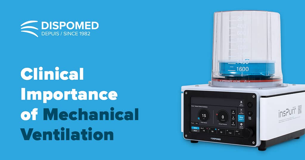
- Driving source- Traditional ventilators utilize high pressure oxygen (40-50 PSI) to drive the bellows and can consume between 10-20 liters per minute of oxygen. Newer ventilators such as the Moduflex InsPurr ventilator eliminate the need for a constant oxygen supply and can even run from a small oxygen concentrator. The turbine-driven technology of the Inspurr not only contributes to financial efficiency but also enhances the overall operational flexibility of your clinic.
- A multi-parameter monitoring system that includes capnography. Capnography is an essential tool for assessing a variety of ventilation patterns.
- An in-circuit pressure alarm is an essential tool for preventing barotrauma caused by increased pressure in the anesthesia circuit. This alarm alerts the anesthetist to accidental pop-off valve closures and signals pressure increases due to circuit or machine malfunctions, such as a kinked endotracheal tube, compromised hoses, or a faulty waste gas system.
What are some important terms that the user needs to be familiar with when using a mechanical ventilator?
MV
A mechanical Ventilator is a machine that generates a controlled flow of gas into a patient’s airways.
IPPV
Intermittent positive pressure ventilation that is maintained only during inspiration (manual or mechanical).
PEEP
Positive End Expiratory Pressure that is used to open small airways during the expiration phase of the breath. This is beneficial for preventing atelectasis, for managing chronic airway disease, lung trauma or pulmonary edema (PEEP: 1-7.5 cm H2O).
PIP
Peak Inspiratory Pressure is inspiratory airway pressure. Normally set between 12-20 cm H2O pressure.
VT
Tidal Volume is a combined full breath (inspired and expired). Calculated 10-20 mls/ kg. Measured with respirometry.
VE
Minute Ventilation is tidal volume x respiratory rate (RR) (Example 200 ml TV x 10 RR= 2 liter VE).
I:E Ratio
Measurement of each breath cycle in the inspiratory and expiratory phase. The duration of each phase will depend on this ratio in conjunction with the overall respiratory rate.
IT
Inspiratory Time is the time to inflate the lungs. This is typically ~ 1 second in the small animal patient.
Ventilatory Rate
Number of cycles per minute. We call this a respiratory rate during spontaneous breathing.
PaO2
Partial pressure of oxygen in arterial blood.
PaCO2
Partial pressure of carbon dioxide in arterial blood.
Respirometry
Quantitative measurement of respiration.
ETCO2
End Tidal caron dioxide. The alveolar measurement of carbon dioxide that is sampled near the endotracheal tube. This is closely correlated to the patients’ arterial CO2 (normal is 35-45 mmHg).
What is hypoxemia and hypoventilation and why do we need to recognize it?
Next to tissue perfusion and blood pressure support, airway maintenance is essential in the anesthesia patient. When a patient is sedated/ premedicated and or anesthetized, the respiratory efforts are decreased, the breathing rate becomes less frequent and chest excursions are more depressed. This impairs the patient’s ability to participate in gas exchange (oxygen for carbon dioxide). When a sedated patient has decreased respiratory efforts, they can become hypoxemic. This is due to hypoventilation (increased carbon dioxide levels). Preoxygenation is important in the pre-operative period as well providing this in the recovery period until normal ventilation returns (ET CO2 35-45 mm Hg and SPO2 of >95%). After induction and intubation, airway support is continued. The need to provide regular ventilation support assistance as it becomes evident if one of the following occurs:
- Hypoxemia is commonly defined as a PaO2 of less than 80 mm or an SPO2 of < 95%
- Hypoventilation is defined as a PCO2 greater than 50 mm Hg or and ETCO2 > 50 mm Hg
- Poor anesthetic depth
Which is better- Manual or Mechanical Ventilation?
Manual and mechanical ventilation in the veterinary patient has always been of importance in veterinary anesthesia as many of our patients have existing conditions or develop complications that require alveolar gas exchange support. Many practices already provide manual breaths to aid in ventilation support. This is essential if a patient becomes apneic or has shallow breathing. Unfortunately, it involves having a dedicated team member that needs to stop what they are doing to be able provide this airway support.
Recently, with improved education and better tools available, more veterinary practices have stepped up their game and now provide controlled ventilation with mechanical units in many of their anesthesia patients. Specifically, cases that have a longer anesthetic events or for patients that may have a physical need such as conditions like obesity, deep chested large breed dogs, brachycephalic patients to name a few. Mechanical ventilators are preferred as they can control airway pressure, volume and rate that is monitoring both by the user and with multiparameter tools (capnography and pulse oximetry). With a mechanical breathing device, this allows the anesthetist to dedicate more time to better monitoring support.
How does a mechanical ventilator help an anesthetized veterinary dentistry patient?
Dental procedures are generally longer anesthetic events due to timing of each process. Procedures may run into hours of anesthesia. Dedicated monitoring is essential for these patients. Pulmonary atelectasis (collapsing of alveoli) occurs shortly after induction of anesthesia and will become more severe with duration. Intermittent positive pressure ventilation will reverse this process. Many patients encountering lengthy dental procedures may have analgesia infusions or will have irregular breathing patterns. A combined balanced anesthesia and analgesic technique as well as a mechanical ventilator will ensure a smooth plane of anesthesia.
Time consuming dental procedures
- Positioning correct monitoring devices
- Preoperative diagnostics (pictures, cone beam CT, oral radiographs, oral exam)
- Full dental prophylaxis sub-gingival, supra-gingival cleaning and polishing
- Treatment- oral surgery, endodontics
- Post surgical assessment (radiographs, CT, pictures)
The dental patient is often positioned with the head lower than the rest of the body. In this position, the abdominal cavity will push on the lungs, increasing breathing resistance. Many dental patients are seniors with underlying co-morbidities. Some are brachycephalic or deep chested. Having a mechanical ventilator will alleviate the stress of poor oxygen/ carbon dioxide exchange.
What is the difference between Pressure versus Volume Controlled ventilation?
Most anesthesia ventilators have volume or pressure control options.
VOLUME option will deliver a preset tidal volume over a given inspiratory time. In a volume-controlled breath, the peak inspired airway pressure (PIP) generated is dependent on the preset tidal volume determined by the operator and the compliance of the respiratory system.
PRESSURE limiting machines can maintain a preset airway pressure for a given inspiratory time. In a pressure-controlled breath, the tidal volume will depend on the preset airway pressure that is determined by the operator and the compliance of the respiratory system.
How can we provide SAFE mechanical ventilation for our anesthetized patients?
The veterinary professional should have a full working knowledge of how the anesthesia machine works and understand what the “normal” vital signs are in the patient (heart rate, blood pressure, oxygen saturation, respiratory rate and character of a normal breath, end tidal caron dioxide levels), as these are often altered by general anesthesia and mechanical ventilation. Include a pressure alarm to monitor safe pressures. There are a few guidelines to consider. Consider the below for an optimal ventilation strategy.
Since patient carbon dioxide and oxygen gas exchange is controlled by the user during manual or mechanical ventilation, observing and maintaining normal values via capnography, pulse oximetry and hands on monitoring is essential. Removal of carbon dioxide is primarily achieved through minute volume. The PIP should fall in the 15-20 cm H2O pressure range. Whether choosing a high ventilation rate with low tidal volume or a low rate with high volume, the effects will be similar.
For time or volume cycled ventilators, calculate the tidal volume first. Observe the patient’s thoracic cavity to assure bilateral chest expansion and maintain a PIP of 15-20 cm H2O pressure. In a pressure cycled ventilator, the inspiratory pressure is set, and chest expansion is observed and adjusted as needed. Observe blood pressure as increasing thoracic cavity expansion may decrease venous return, which may or may not impact mean arterial pressures. The inspiratory time should always be less than the expiratory time as this will allow full expansion of the thoracic cavity, move gas in and out of the alveoli and lastly, allow enough time for recoiling of the lungs. Some mechanical ventilators have internal respirometry that measures each patient’s ventilation needs. This type of ventilator has a wider safety range. When weaning a patient from the mechanical ventilator, allow the patient to build of carbon dioxide to trigger normal ventilation as this is determined by the respiratory center in the brain. You can decrease the rate and volume or continue to manually support ventilation needs until the patient has returned to normal breathing volume and . Always observe carbon dioxide parameters until patient is regaining consciousness as well as blood oxygen via a pulse oximeter and pink color of mucosa.
How do I choose a SAFE mechanical ventilator?
Many ventilators have a bellows style construction that may be driven pneumatically or via a piston or turbine. There also may be microprocessors that assist with the drive. With pneumatic driven ventilators, oxygen consumption is high. Other features include safety pressure alarms and relief valves. An anesthesia machine pressure alarm should always be included when a ventilator is in use to prevent accidental pop off valve closure. There are also pop-off valves that have unique safety features which prevent the user from closing the valve when a ventilator is in use. The pressure occlusion occurs when a cap is in place. Newer ventilators such as the Inspurr, that is turbine driven use zero liters per minute of oxygen to drive the bellows, is quiet and safer to use due to the internal respirometry that will measure the patient’s lung volume assuring an accurate positive pressure breath.
References
Cooley, Kristen G. & Johnson, Rebecca A. (2018). Veterinary Anesthetic and Monitoring Equipment. Hoboken: Wiley and Sons
Dorsch & Dorsch, 5th ED (2007). Understanding Anesthesia Equipment. Baltimore: Lippincott Williams & Wilkins
Seymour, Chris & Duke-Novakovski, Tanya. (2016). BSAVA Canine and Feline Anaesthesia and Analgesia, 3rd ED. Gloucester: BSAVA