Table of Contents
In August of 2018, the National Association of Veterinary Technicians in America (NAVTA) announced the creation of the sixteenth veterinary technician specialty: Veterinary Technician Specialist in Diagnostic Imaging (VTS-DI).
This specialty joins a list of 15 other veterinary technician specialties:
- Emergency and Critical Care
- Dentistry
- Internal Medicine
- Laboratory Animal Medicine
- Anesthesia and Analgesia
- Veterinary Behavior
- Clinical Pathology
- Clinical Practice
- Dermatology
- Equine Veterinary Nursing
- Physical Rehabilitation
- Nutrition
- Ophthalmology
- Surgery
- Zoological Medicine
The Veterinary Technician Specialist in Diagnostic Imaging title is intended to recognize those veterinary technicians specializing in the field of advanced diagnostic imaging. Here is an overview of their primary responsibilities and career perspectives.
Veterinary medical imaging: techniques and applications
Veterinary diagnostic imaging technicians are typically employed by veterinary specialty practices or colleges of veterinary medicine.
A VTS-DI works under the supervision of a veterinarian, typically a veterinary radiologist. In some cases, a VTS-DI may also assist internal medicine specialists, surgeons, neurologists, or other specialists using advanced diagnostic imaging modalities.
Common techniques employed by veterinary diagnostic imaging technicians include:
- Digital radiographs
In this type of radiographic imaging, digital x-ray sensors are used instead of film. There are two distinct types of digital radiographic systems. In digital radiography (DR), the digital sensors are embedded directly in the table of the x-ray machine; when an x-ray image is taken, the image is automatically transferred to a computer monitor for viewing.
In computed radiography (CR), a cassette system is used; the cassettes contain a phosphor imaging plate instead of traditional film. This cassette is inserted into a computerized reader, then the image appears on a computer screen. Each system has both advantages and disadvantages. Veterinary diagnostic imaging technicians may work with either of these systems.
- Fluoroscopy
This type of medical imaging shows a continuous, real-time radiographic image on a monitor, almost like a movie. Fluoroscopy is often used for surgical and cardiac procedures, as it allows the surgeon to visualize the internal location of catheters and other instruments as they are inserted into the body.
- Computed Tomography (CT)
This computerized radiographic technique is used for detailed scans of internal structures, including bones, soft tissues, blood vessels, and internal organs. CT images are typically viewed as “bread slices” through the patient, though they can be reconstructed into 3D images for visualization.
- Magnetic Resonance Imaging (MRI)
This diagnostic imaging modality measures the response of internal tissues to radio waves transmitted while the body is in a strong magnetic field. MRI imaging is similar to CT in that it allows detailed visualization of internal structures, but MRI can often offer greater detail than that provided with CT imaging.
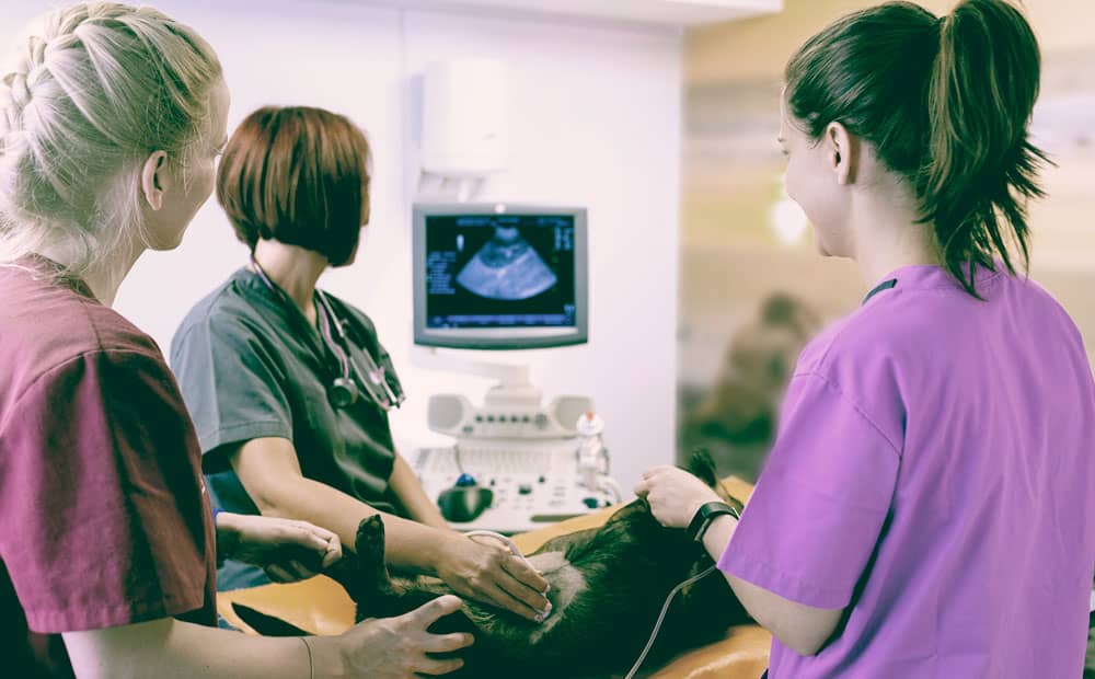
- Ultrasound
This type of imaging uses the reflection of sound waves to image internal structures. Ultrasound imaging is less detailed than CT or MRI, but is also significantly less expensive and has the benefit of allowing visualization of real-time movement and function of internal structures.
- Nuclear medicine imaging
Radioactive tracers are injected or administered to a patient, then nuclear medicine imaging is used to visualize the uptake of these tracers by various body organs. Nuclear imaging is often used to evaluate the thyroid gland, though it also has other applications in veterinary medicine.
What is hands-free radiology?
Veterinary diagnostic imaging technicians are expected to be well-versed in the application of hands-free radiology techniques. These techniques promote radiation safety and OSHA compliance by decreasing radiation exposure among members of the veterinary team.
One component of hands-free radiology is sedation, which is recommended in many cases. This allows patients to be positioned for radiographs more easily, without requiring a technician to stand near the primary beam to restrain them.
Other components of hands-free radiology include the use of tools or materials designed to promote proper patient positioning without a need for manual restraint. Foam wedges, sandbags, elastic straps, and V-trays are often used as positioning tools in hands-free radiology.
A day in the life of a veterinary diagnostic imaging technician
As a veterinary diagnostic imaging technician, you will likely be employed by a private specialty practice, corporate specialty practice, or college of veterinary medicine. You will work under the supervision of radiologists or other specialists, assisting with diagnostic imaging of patients. If you work in a college of veterinary medicine, you may also find yourself teaching veterinary students.
Your day will likely start early, around 8 am. Depending on the size of your facility and how many diagnostic imaging technicians are on your team, you may be assigned to a specific area of the radiology department or you might find yourself assisting with a wide variety of cases. Regardless of which of these situations applies, you will likely be assigned to a list of specific radiology cases for the day and expected to follow these cases from start to finish.
Some of your cases may involve taking digital radiographs on small or large animal patients. Small animal digital radiology is typically performed using a stationary DR or CR system. Large animal patients may be imaged with stationary systems or with portable digital x-ray systems. After obtaining images of your patient, you will be expected to review these images for quality control, ensuring that positioning and technique are appropriate so that the radiologist (or other specialist) can interpret the images.
You may also work with ultrasound cases. Veterinary ultrasound units vary significantly, depending on practitioner needs and anticipated uses. An ultrasound unit is typically accompanied by a number of transducers (probes), each used for different types of ultrasound imaging.
As a veterinary diagnostic imaging technician, you will be expected to prepare the ultrasound with appropriate transducers, assist the veterinarian performing the ultrasound, and possibly perform some ultrasounds independently (collecting images that will be interpreted by the radiologist).
Advanced imaging studies, such as CT, MRI, and fluoroscopy, are typically performed while pets are anesthetized. If you are assigned to one of these cases, your responsibilities will depend on the overall staffing and workflow of your practice. In some practices, you may find yourself responsible for anesthetizing the patient and performing the patient’s imaging. In other practices, you may have an anesthesia team to handle anesthesia induction, monitoring, and recovery, allowing you to focus on obtaining the imaging studies.
Because MRI imaging uses powerful magnets, specialized MRI-compatible veterinary anesthesia machines must be used in the MRI suite to prevent injury to machines, patients, and members of the veterinary team.


Application and training process for the VTS-DI
In order to become recognized as a VTS-DI, candidates must apply to the Academy of Veterinary Technicians in Diagnostic Imaging (AVTDI).
Candidates must submit extensive documentation attesting to their radiographic skills, along with a case log documenting specific experiences in general radiology, advanced imaging/contrast, and nursing and pharmacology. Each case must be signed off on by a veterinary radiologist (or other veterinary specialist) or an existing member of the AVTDI.
If this application is approved, candidates will be invited to sit for the VTS-DI certifying exam. This examination will include multiple choice, written, and practical test items covering a variety of topics in veterinary imaging. Sample topics include:
- Radiation safety
- Radiographic positioning for various species
- Fluoroscopy
- Image quality control
- Pharmacology
- Medical records
Candidates will also be tested on their knowledge of CT, MRI, nuclear medicine, and ultrasound.
Any individual that is approved to take the certifying exam and obtains a passing score will be awarded the title Veterinary Technician Specialist – Diagnostic Imaging. Once awarded, this title will remain in effect for five years.
Salary and employment prospects
As more and more pet owners demonstrate a willingness to seek advanced medical care, including advanced imaging techniques such as CT and MRI, the need for veterinary diagnostic imaging technicians is expected to increase.
Because the VTS-DI is a relatively new specialty, little information exists regarding anticipated salary ranges. In general, full-time veterinary technician specialists earn approximately $45,000 per year, so a VTS-DI would be expected to have earnings in this range.
Sources and additional reading
- 2018. Diagnostic Imaging Veterinary Technician Specialty Recognized by NAVTA. https://cdn.ymaws.com/www.navta.net/resource/resmgr/news_related/Press_Release_Diagnostic_Ima.pdf
- American College of Veterinary Radiology. Veterinary Imaging or Radiation Oncology Technologist. https://www.acvr.org/page/veterinary-imaging-or-radiation-oncology-technologist
- Hands Free X-rays. http://handsfreexrays.com
- The Academy of Veterinary Technicians in Diagnostic Imaging. http://avtdi.org/
- Kenwright, M. 2015. What specialized veterinary technicians bring to the table. Available at: http://veterinarymedicine.dvm360.com/what-specialized-veterinary-technicians-bring-table
About the author


